
Figure 4.
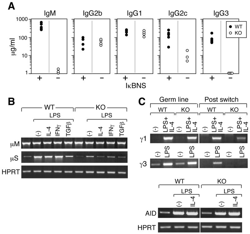
IκBNS KO mice manifest B cell defects in serum immunoglobulin levels and IgG3 switch recombination. A) Basal immunoglobulin isotype levels in the sera from naïve IκBNS KO (-) mice and age-matched WT/HET mice (+) were measured by ELISA. Immunoglobulin levels less than 0.1 μg/ml were plotted on the base line. The serum IgA levels of IκBNS KO mice were similar to that of C57BL/6 mice (data not shown). p<0.05 for IgG2c; p<0.01 for IgM and IgG3. All mice are on a C57BL/6 background: 1 mouse was WT, 4 mice were IκBNS heterozygous and 4 mice were IκBNS KO. B) Splenic B cells (CD43-) from WT and IκBNS KO mice were stimulated in vitro with LPS (5 μg/ml) alone or LPS plus cytokines (10 ng/ml) as indicated for 4 days. Semi-quantitative RT-PCR analysis of μM, μS and HPRT mRNA was performed. C) Germline and post-switch transcripts for IgG1 and IgG3 were assessed in cDNA prepared from cells cultured in the presence and absence of LPS alone or in combination with with IL-4, respectively. Lower panels: AID and HPRT expression before and after stimulation with LPS or LPS plus IL-4 were examined. AID expression confirms that switch recombination processes have initiated and HPRT expression confirms equivalent loading of RNA in each sample. For B and C, two independent experiments were performed and similar results were obtained.