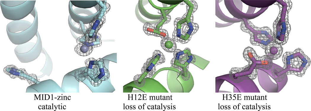
Figure 5.
Crystallographic evidence for the catalytic mechanism. A) The MID1-zinc crystal structure (PDB code 3V1C) reveals a cleft and open zinc coordination site. B) The H12E mutation (PDB code 3V1E) and C) the H35E point mutation (PDB code 3V1F) close the cleft and complete the four-coordination of zinc, and these mutants demonstrate loss of catalytic activity.

