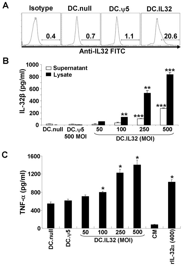
Figure 1. Expression and bioactivity of hIL-32β in mouse CD11c+ DCs.
When compared to control DCs (DC.null and DC.ψ5), DC.IL-32 expressed higher intracellular expression levels of IL-32β protein as determined by flow cytometry (panel A; MOI = 250) and ELISA (panel B; MOI ranging from 50–500) as outlined in Materials and Methods. For the ELISA assays (B), both DC culture supernatants and DC lysates were analyzed for IL-32β concentration. In C, cell-free supernatants were harvested 48h after DC infection with no adenovirus, Ad.ψ5 (MOI = 500) or Ad.IL32β (at MOI ranging from 50–500), and then used to supplement cultures of the Raw 264.7 murine macrophage cell line. After 18h, supernatants were recovered and analyzed for TNF-α content by ELISA. Recombinant IL-32α (400 pg/ml) was used as positive control for induction of TNF-α from Raw 264.7 cells. * p < 0.05; ** p < 0.01; *** p < 0.001.

