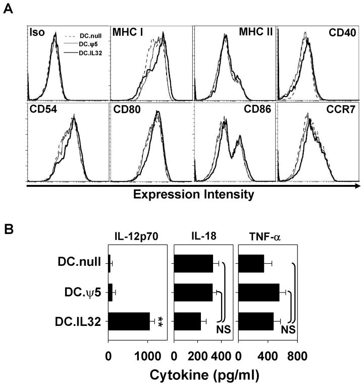
Figure 2. Ectopic expression of IL-32β enhances the immunostimulatory phenotype of DC.
DC.IL32 (MOI = 250) or control DC (DC.null or DC.ψ5) were prepared as described in Materials and Methods. Forty-eight hours after adenoviral infection, DC were harvested and analyzed by flow cytometry for cell surface expression of MHC and costimulatory molecules, as well as, CCR7 required for DC migration to secondary lymphoid tissues (A). In B., cell-free supernatants were recovered from the individual DC cohorts 24h after stimulation with CD40L+J558 cells (as described in Materials and Methods) and analyzed for the indicated cytokines by specific ELISA. Results are reported as the mean ± SD of triplicate determinations. NS = not significant; **p < 0.01.

