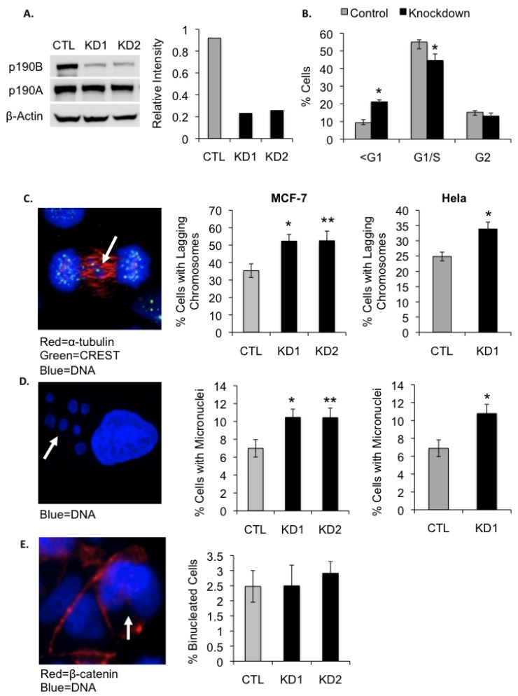
Figure 2.
P190B deficiency induces lagging chromosomes and micronucleation in MCF-7 and Hela cells. Western blot and graph representing normalized densitometry values show an approximately 75% reduction in p190B protein levels in MCF-7 cells transfected with p190B-targeting siRNA compared to cells transfected with control non-targeting siRNA. siRNA targeting P190B did not affect the expression levels of the closely related p190A RhoGAP as determined by Western blotting (A). The percentages of control and p190B knockdown cells in different stages of the cell cycle as determined by flow cytometry are graphed, * p < 0.001 (B). A representative confocal image of an anaphase MCF-7 cell immunostained with an antibody against α-tubulin and CREST anti-serum is shown. Arrow indicates a lagging chromosome. The percentages of control and p190B deficient MCF-7 and Hela cells with lagging chromosomes at anaphase are graphed, * p = 0.012, ** p = 0.03 (C). A representative confocal image of an interphase MCF-7 cell stained with DAPI is shown. Arrow indicates micronuclei. The percentages of control and p190B knockdown MCF-7 and Hela cells with micronuclei are graphed. For MCF-7 * p = 0.027, ** p = 0.04 and for Hela, * p = 0.019 (D). A representative image of MCF-7 cells immunostained with an antibody against β-catenin is shown. DAPI was used to stain DNA. Arrow indicates a binucleated cell. The percentages of binucleated control and p190B deficient MCF-7 cells are graphed (E).

