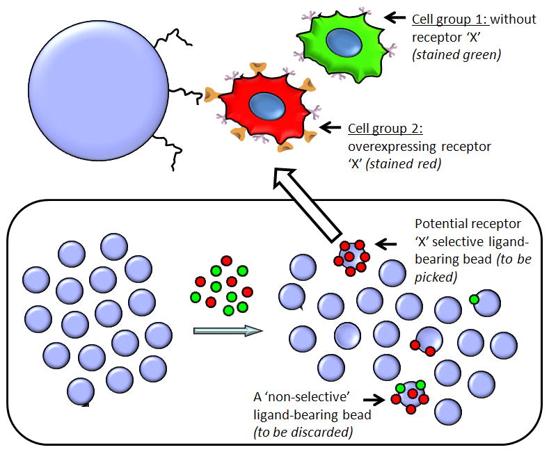
Figure 1.

Schematic representation of the on-bead two-color (OBTC) assay for the identification of peptoid ligands for receptor ‘X’. Blue circles represent peptoid library beads. The single bead shown on top of the figure displays a red stained receptor ‘X’ overexpressing cell binding to a peptoid molecule on the bead through the specific interaction of the receptor ‘X’. Since this peptoid is highly specific and only binds to receptor ‘X’, it does not recognize any cell surface molecule on the parent cells (green stained) and hence no green cells attached to this bead. Schematic representation of the bottom panel depicts the actual equilibration of two stained cell types (red and green in 1:1 mixture) with OBOC library beads. A potential ‘hit’ (with only red cells) and a non specific peptoid carrying bead (with both cell types) is schematically shown in the bottom right.
