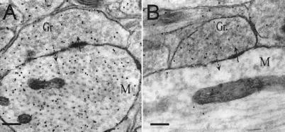
Figure 2.
Electron micrographs showing immunogold labeling for glutamate (Glu-LI, A) or GABA (GABA-LI, B) in reciprocal synapses between GC spines (Gr) and MC dendrites (M) in the EPL. The mitral to granule contact is asymmetric (solid arrow) and the granule to mitral contact is symmetric (dotted arrow). Glu-LI is found in MC and GC (A). GABA-LI is localized to the GC (B). Particle densities (particles/μm2) for Glu-LI are 63.1 ± 5.8 over M and 86.5 ± 5.2 over Gr (mean ± SE). Particle densities for GABA-LI are 9.7 ± 3.3 over M and 30.1 ± 2.8 over Gr. Background noise, assessed over empty resin, was of 4 ± 2.3 particles/μm2 for Glu-LI and 3.1 ± 0.2 particles/μm2 for GABA-LI. (Scale bars: 240 nm.) Electron micrographs were obtained from a Phillips CM120 electron microscope at the Université Claude Bernard-Lyon.

