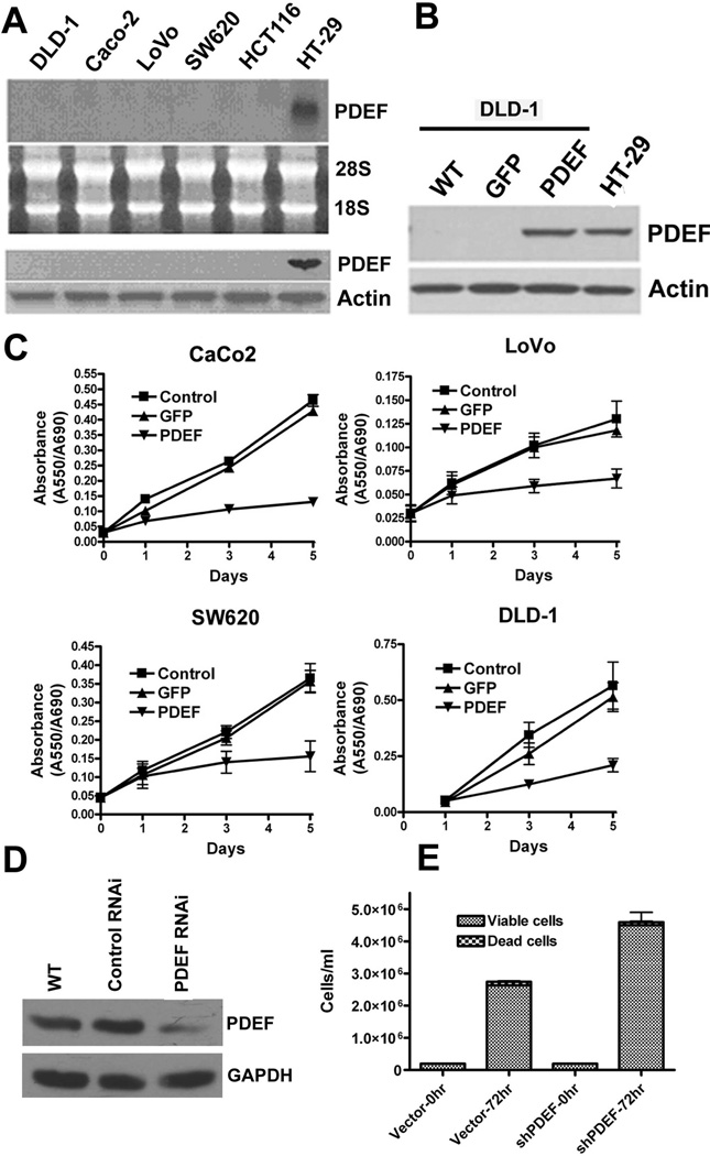
Figure 1. PDEF is lost in invasive colon cancer cell lines and re-expression of PDEF leads to growth changes in colon cancer cells.
(A) Northern and Western blot analyses of RNA and protein prepared from the indicated colon cancer cell lines. Ethidium bromide staining of 28S and 18S rRNA or actin levels are provided as a controls. (B) Western blot analysis of PDEF expression level after infection of DLD-1 colon cells and endogenous expression level observed in HT-29 cells. Actin levels provided as a control. (C) CaCo-2, LoVo-2, SW680 and DLD-1 cells were untreated (control), or infected with either Ad-GFP or Ad-PDEF and cellular growth was monitored by MTT assay for 5 days. (D) Western blot analysis of PDEF protein expression in HT-29 colon cancer cells and in HT-29 expressing vector (RNAi control) and short hairpin RNA (shRNA) PDEF. Actin levels provided as a control. (E) Number of HT-29 colon cancer cells expressing vector alone or PDEF shRNA measured after 72 hours (p<0.05).

