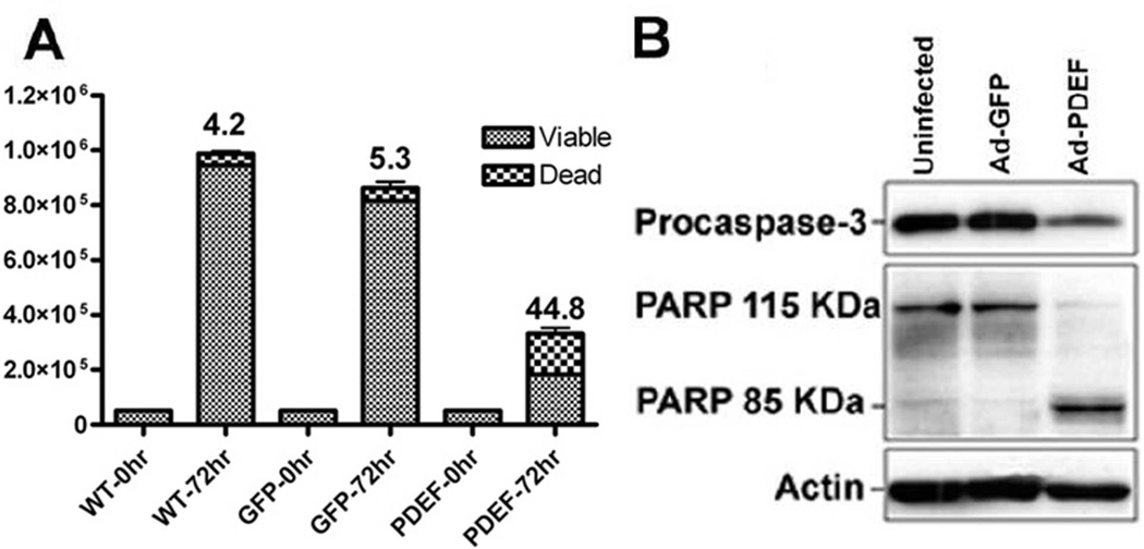
Figure 3. PDEF expression increases apoptosis.
(A) Quantitation of trypan blue cell viability assays of parental (uninfected) DLD-1 cells, or cells infected with Ad-GFP or Ad-PDEF. The columns represent the average values for apoptotic index, the percentage of apoptotic vs. total cell number. Results are statistically significant with a p-value <0.05. (B) Western blot analysis of procaspase 3 and PARP expression levels in DLD-1 cells infected with Ad-GFP or Ad-PDEF. Actin levels provided as a control.

