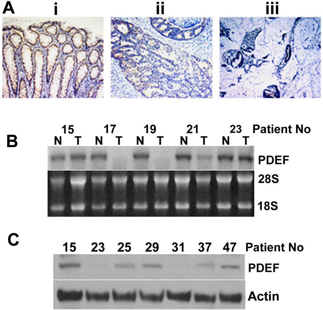
Figure 5. PDEF expression is reduced in colon cancer tissue.
(A) Immunohistochemical analysis of PDEF expression in colon tissue: (i) non-tumor, (ii) tumor and (iii) invasive mucinous portion of tumor. Panels are from the same section. (Magnification, X400). Data representative of 5 of 6 cases. (B) Northern blot analysis for PDEF mRNA in a series of matched tumor (T) and non-tumor (N) tissues from the same patient (indicated by numbers). Data representative of 25 matched tumor/non-tumor samples. Ethidium bromide staining of 28S and 18S rRNA provided as control. (C) Western blot analysis of PDEF protein in 7 tumors that retain PDEF mRNA expression. Actin levels provided as a control.

