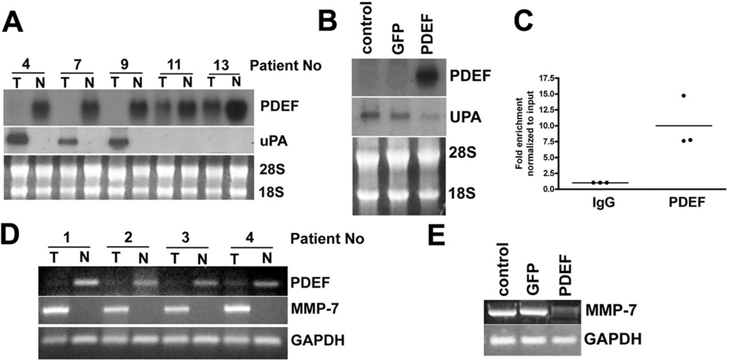
Figure 6. PDEF loss alters the expression of the cancer associated genes, uPA and MMP-7.
(A) Northern blot analysis of RNA prepared from tumor tissue and matched adjacent non-tumor tissues. 28S and 18S RNA provided as control for loading and RNA integrity. Data is representative of 25 tumor and non-tumor pairs examined. (B) Northern blot analysis of RNA prepared from parental DLD-1 cells and DLD-1 cells infected with Ad-GFP or Ad-PDEF. Ethidium bromide staining of 28S and 18S provided as a control. (C) Chromatin immuno-precipitation (CHIP) analysis using chromatin prepared from the HT-29 cell line. Q-PCR analysis using primers for the uPA promoter and chromatin immunoprecipitation with a PDEF-specific antibody, compared to IgG control antibody. (D) RT-PCR analysis MMP-7 mRNA in RNA prepared from tumor tissue and matched adjacent non-tumor tissues. GAPDH RT-PCR provided as a control. Data representative of 25 matched samples. (E) RT-PCR analyses of MMP-7 mRNA in RNA prepared from uninfected DLD-1 cells and cells infected with Ad-GFP or Ad-PDEF. GAPDH provided as control.

