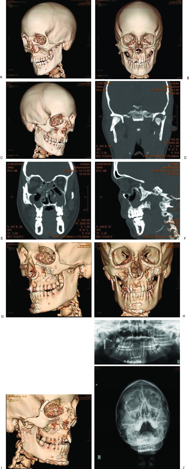
Figure 1.
Selected preoperative radiographic CT images in right (A) oblique, (B) frontal, and (C) left oblique view, of a female patient 22 years of age with complex panfacial fractures of midface and mandible. Image (D) shows a coronal cut of dislocated fractures of the right mandibular condyle and left mandibular head. Image (E) depicts dislocated Le Fort I, zygoma and orbital floor fractures. Image (F) reveals the extent of the orbital floor fracture in the anterior posterior direction. Selected postoperative radiographic CT images in (G) right oblique, (H) frontal, and (I) left oblique view, and (J) conventional radiographs, show the great current options of matrix plates and screws for treating CMF Trauma.

