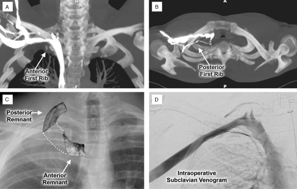
Figure 3.
Paraclavicular approach for reoperative treatment of venous thoracic outlet syndrome. Images from an active young man who had developed right-sided subclavian vein (SCV) effort thrombosis and was treated by thrombolysis and first rib resection. Over the following year he continued to experience exertional right upper extremity swelling and pain, despite several attempts at SCV balloon angioplasty. (A) Maximal intensity projection coronal (MIP) images from a contrast-enhanced computed tomography study demonstrating a persistent focal stenosis in the proximal right SCV during arm elevation, located adjacent to a small remnant of the anterior first rib (arrow). (B) Axial MIP images demonstrating expanded collateral venous pathways passing through the thoracic outlet adjacent to a remnant of the posterior first rib (arrow). (C) Operative specimens of the anterior and posterior first rib remnants removed during paraclavicular decompression, superimposed on the plain chest radiograph (arrows). (D) Intraoperative contrast venogram obtained following first rib resection and external venolysis, demonstrating a widely patent SCV with minimal filling of collateral venous pathways.

