Figure 4.
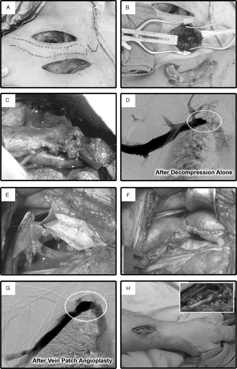
Surgical management of subclavian vein (SCV) effort thrombosis by paraclavicular thoracic outlet decompression and direct venous reconstruction. (A) Supraclavicular and infraclavicular incision locations for thoracic outlet decompression. (B) Following scalenectomy and division of the posterior first rib through the supraclavicular exposure, the medial first rib is exposed and divided at the level of the sternum through the infraclavicular exposure, and the entire first rib is removed as a single specimen. (C) Following resection of the subclavius muscle and exposure of the axillary vein through the infraclavicular incision, the proximal SCV is exposed through the supraclavicular incision. The SCV is dissected to its junction with the internal jugular and innominate veins, and fibrous scar tissue surrounding the SCV is resected. A focal area of persistent fibrosis in the proximal SCV is present despite external venolysis. (D) Intraoperative venogram following the decompression portion of the operation, demonstrating a residual high-grade obstruction of the proximal SCV (oval). (E) A longitudinal venotomy is created in the proximal SCV, extending through the site of the focal stenosis. The luminal surface of the SCV is free of thrombus or ulceration. (F) Construction of a vein patch angioplasty of the SCV, extending into the anterolateral aspect of the innominate vein. (G) Completion intraoperative venogram following vein patch angioplasty, demonstrating restoration of a widely patent SCV with no filling of venous collaterals. (H) Construction of an adjunctive radiocephalic arteriovenous fistula at the right wrist, which will be subsequently ligated 12 weeks after the primary operation.

