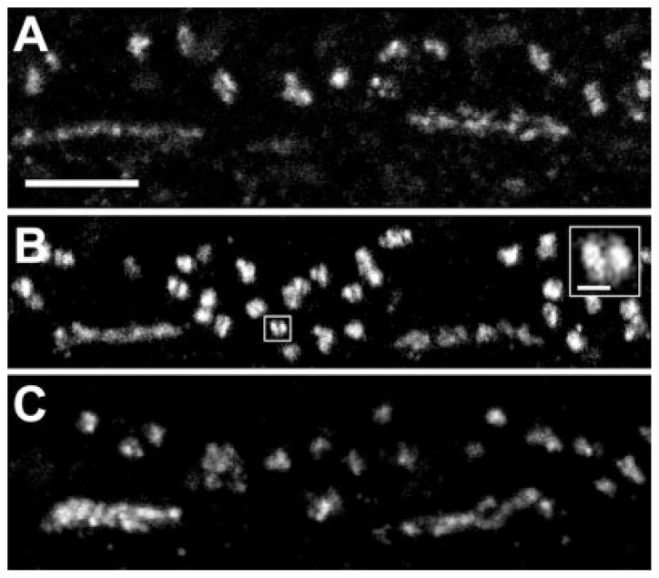
Fig. 2.
HR3 in the OPL of macaque (A,B) and baboon (C) retinas. The pattern of labeling in the OPL was identical with the two antibodies raised against HR3, HR3C (A) and HR3N (B). Clusters of small, HR3C-immunoreactive (IR) puncta formed a band at the bases of cone pedicles. Numerous larger puncta sclerad to the cone pedicles were associated with rod spherules. Some of the large puncta were composed of two smaller puncta in close apposition (inset). C: By using an antibody to HR3C, an identical pattern of labeling was found in baboon retina. Scale bars = 5 μm in A–C; 1 μm in inset.

