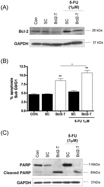
Figure 6. Repression of Bcl-2 potentiates 5-FU-induced apoptosis in metastatic prostate cancer cells.
(A) Immunoblot presenting the expression level of the anti-apoptotic protein Bcl-2 in PC3 cells transfected with a non-targeting oligonucleotide (Sc) or a gene-targeted oligonucleotide (Bcl2-T). All oligonucleotides were used at a final concentration of 50 nM. Expression of Bcl-2 is presented in the absence or presence of 1 µM 5-FU. (B) Bar graph presenting the apoptotic cell population determined by flow cytometry analysis in PC3 cells transfected with a non-targeting oligonucleotide (Sc) or a gene-targeted oligonucleotide (Bcl2-T) at a final concentration of 50 nM, in the absence or presence of 1 µM 5-FU. Statistically-significant differences in the apoptosis levels were determined by conducting two-tailed Student's t-test analysis; *, P<0.05; **, p<0.01. (C) Immunoblot presenting the expression and cleavage product of PARP in PC3 cells transfected with a non-targeting oligonucleotide (Sc) or a gene-targeted oligonucleotide (Bcl2-T), used at a final concentration of 50 nM, in the absence or presence of 1 µM 5-FU. Equal protein loading in immunoblots shown in (A) and (C) was confirmed by re-probing the membranes for expression of GAPDH.

