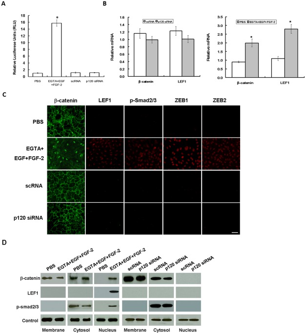
Figure 3. Activation of Wnt and Smad/ZEB signaling by EGTA+EGF+FGF-1 but not p120 knockdown.
ARPE-19 cells cultured to day 7 post-confluence were treated with PBS or 1 mM EGTA plus 10 ng/ml EGF and 20 ng/ml FGF-2 for 1 day with or without 5 ng/ml XAV939, or transfected with 100 nM scRNA or p120 siRNA for 2 days. (A) The TCF/LEF1 promoter activity was silent in PBS, scRNA, or p120 siRNA, dramatically elevated to nearly 16-fold by EGTA+EGF+FGF-2 (P<0.05), but completely abolished by XAV939 (not shown). (B) qRT-PCR showed that β-catenin and LEF1 transcripts were up-regulated 2.2- and 2.5-fold, respectively (P<0.05), in EGTA+EGF+FGF-2, but remained unchanged in p120 siRNA (P>0.05). (C) Immunostaining reveal increased nuclear staining of β-catenin, LEF1, p-Smad2/3, ZEB1, and ZEB2 in EGTA+EGF+FGF-2 but not p120 siRNA. Scale bar indicates 100 μm. (D) Western blot analysis confirmed that the β-catenin level decreased in the membranous extract but increased in the nuclear extract, while the LEF1 level increased in the nuclear extract in EGTA+EGF+FGF-2, while there was no such change by p120 siRNA. Connexin43, α-tubulin, and histone serve as the loading control for membranous, cytosolic, and nuclear extracts, respectively.

