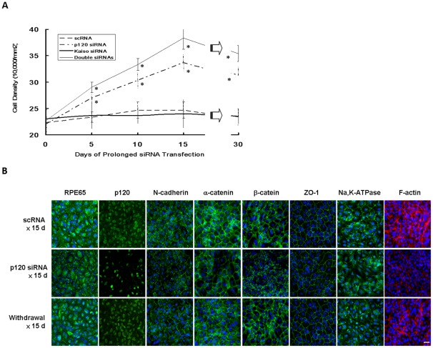
Figure 6. Compact ARPE monolayer with a normal phenotype without EMT after prolonged p120 knockdown.
ARPE-19 cells cultured to day 7 post-confluence were transfected with 40 nM scRNA or siRNA to p120, Kaiso, or both for every 5 days until day 15, followed by withdraw for another 15 days. (A) The baseline cell density was comparable before (day 0) transfection, and reached a plateau of 24.7±2.5×10⁴/mm2 (n = 3) and 24.0±2.7×10⁴/mm2 (n = 3, P>0.05) in scRNA and Kaiso siRNA, respectively. In contrast, it increased to 33.7±1.5×10⁴/mm2 (n = 3, P<0.05) and 38.3±2.1×10⁴/mm2 (n = 3, P<0.01), respectively, in p120 siRNA or both siRNAs on day 15. After withdrawal, the final density of all groups slightly decreased on day 30, when same cell morphology was noted as day 15 but dome-shaped areas disappeared (* P<0.05). (B) Immunostaining revealed decreased peri-membranous staining of F-actin and mildly decreased junctional staining of p120, N-cadherin, α-catenin, β-catenin, ZO-1, membranous and junctional staining of RPE65, membranous staining of Na,K-ATPase after p120 siRNA transfection for 15 days. Normal expression patterns were regained after withdrawal for 15 days. Scale bar indicates 100 μm.

