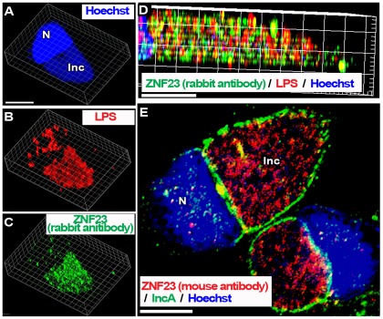
Figure 7. The nuclear ZNF23 protein is mobilized into the lumen of the Ct inclusion.
HeLa cells were grown on coverslips and infected in cell culture medium without antibiotics and cycloheximide. Cells were infected at a MOI of 1 with Ct strain L2 (panel A to D) and with D (panel E) and were fixed 36 hr post-infection. DNA was detected with the Hoechst dye (blue). In cells infected with L2, the bacteria were detected with a mouse antibody against LPS (red, panel B and D) and the ZNF23 protein was detected with a rabbit antibody (green, panel C and D). Z stack images were deconvolved and single channel Surpass 3D view of a representative infected cell is shown in panel A to C. Cropped longitudinal view of the Surpass (x, y, z) view of the merged image is shown in panel D. In Panel E, the inclusion membrane was detected with a rabbit antibody against the bacterial IncA protein (green) and the ZNF23 protein was detected with a mouse antibody (red), and the merged image with Hoechst staining is shown. Images were taken with a Zeiss LSM710 confocal microscope at 63x magnification. The bars in the panels represent 10 µm. Nuclei and inclusion are indicated with N and Inc, respectively.

