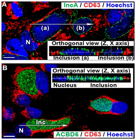
Figure 9. CD63 is not localized in the Ct inclusion of infected HEp-2 cells.
HEp-2 cells were grown on coverslips and infected with Ct strain D at a MOI of 1 in cell culture medium without antibiotics and cycloheximide. Cells were fixed 44 hr post-infection. In panel A and B, CD63 protein was detected with a mouse monoclonal antibody (red). In panel A, the inclusion membrane was stained with a rabbit antibody against IncA (green), DNA was detected with the Hoechst dye (blue), and the merged image is shown. In panel B, the bacteria were only detected by Hoechst DNA staining and ACBD6 was detected with a rabbit antibody (green), and the merged image is shown. Signal distribution of the orthogonal (z, x) slice of deconvolved Z stack images, indicated by the arrow on the respective merged image, are displayed in the inset of each panels. Images were taken with a Zeiss LSM710 confocal microscope at 43x magnification. The bars in panels represent 10 µm. Nuclei and inclusion are indicated with N and Inc, respectively. Note that CD63 signal was not detected in the inclusions (panel A) and did not co-localize with ABCD6 protein, which was detected inside the inclusions (panel B).

