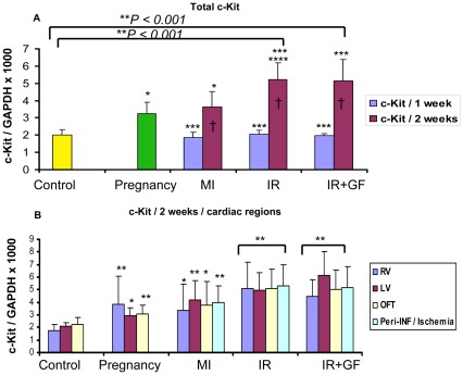
Figure 2. Quantitative RT-PCR showing the time-dependent distribution of c-Kit mRNA in the different groups.
The c-Kit mRNA expression in the different groups is related to the expression of GAPDH. Fig A shows the mean of the whole heart c-Kit mRNA expression in each group. In fig B, the distribution of the c-Kit mRNA between each region at 2 weeks is demonstrated. Number of animals is 7–10 per group (see flow chart in Fig 1). In figure; MI: myocardial Infarction; IR: ischemia-reperfusion; IR+GF: ischemia-reperfusion+ growth factors. RV: Right ventricle, LV: Left ventricle, OFT: Outflow tract and Peri-INF/Ischemia: Peri-infarction and Ischemia region respectively. Data is presented as mean ± SD. *P<0.05 vs. Control group, ** P<0.001 vs. Control group, ***P<0.05 vs. pregnancy group, ****P<0.05 vs. MI (2 weeks) group and † P<0.05 1 week vs. 2 weeks. In fig B, each region is related to corresponding region of the control, pregnancy, myocardial infarction and ischemia-reperfusion w/o growth factors.

