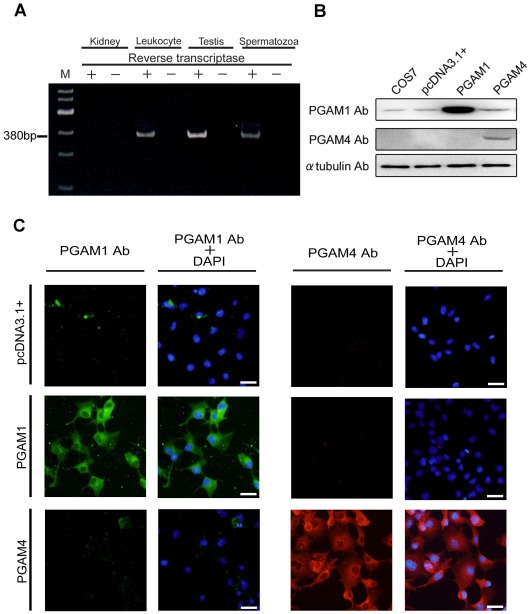
Figure 1. Gene expression analysis of PGAM1 and PGAM4.
(A) PGAM4 mRNA detection in human leukocytes, testes and ejaculated spermatozoa. Total RNA samples from leukocytes, testes and spermatozoa were subjected to RT–PCR analysis. RT–PCR without reverse transcriptase confirmed the absence of DNA contamination after DNase treatment. M, 100-bp ladder DNA marker. (B) Western blot analysis of transfected COS7 cell lysates to determine the specificity of the anti-PGAM1 and anti-PGAM4 antibodies for each antigen. Approximately 8 µg of protein from the transfected cell lysate was loaded. COS7 and pcDNA3.1+ indicate proteins from untransfected cells and cells that were transfected with the empty pcDNA3.1+ vector, respectively. (C) Immunofluorescence analysis of COS7 cells engineered to overexpress PGAM1 or PGAM4. pcDNA3.1+ indicates cells that were transfected with pcDNA3.1+ as a negative control. The nuclei were counterstained with DAPI (blue). PGAM1 Ab, PGAM1-specific antibody; PGAM4 Ab, PGAM4-specific antibody.

