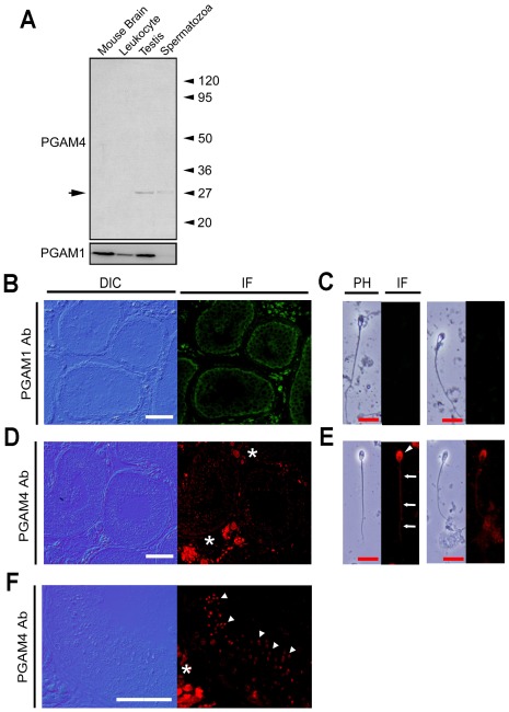
Figure 2. Gene expression and localization of PGAM1 and PGAM4.
(A) Western blot analysis of total protein from leukocytes, testes, spermatozoa and transfected cell lysates. Mouse brain lysate was loaded as a positive control for PGAM1. (B–F) Immunohistochemical observations. Immunofluorescence in the testis was measured using the anti-PGAM1 (B) and anti-PGAM4 (D, F) antibodies. (F) High-power field of D. Immunofluorescence in the spermatozoa was measured using the anti-PGAM1 (C) and anti-PGAM4 (E) antibodies. PGAM4 protein was detected postmeiosis in spermatids and spermatozoa (white arrowhead) and was localized to the principal piece (white arrow) and acrosome (white arrowhead) in ejaculated spermatozoa. Stars indicate nonspecific fluorescence signals from the secondary antibody in interstitial Leydig cells, because negative control experiments without primary antibodies showed the signals in the interstitial area (data not shown). White bars = 20 µm; red bars = 10 µm.

