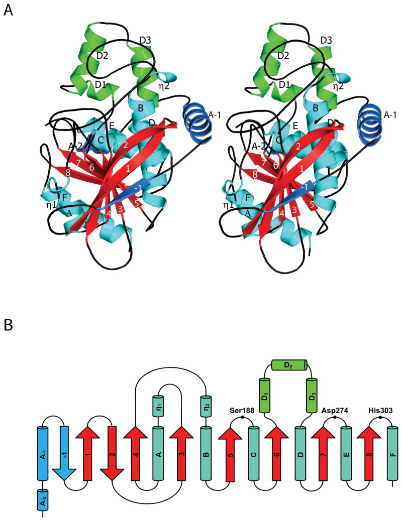
Figure 2.
Overall fold and topology of TM0077. (A) Stereo view of a TM0077 protomer. The β-strands are labeled numerically (-1 to 8) with the core strands in red, α-helices are labeled alphabetically (A-2 to F) and 310-helices are labeled with an Eta (η1 and η2) with the core helices in cyan. The three-helix insertion after β6 is colored green and the N-terminal extension is colored sky blue. The figure was generated using Pymol 31. (B) Topology diagram of TM0077, with the helices displayed as cylinders and the strands displayed as arrows following the color and label scheme of (A). The location of residues forming the catalytic triad is also indicated.

