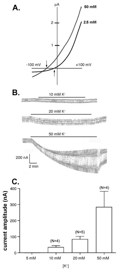
Figure 1. Elevated extracellular K+ activates Panx1 channels in oocytes.
(A) Example of currents recorded from mPanx1 expressing oocytes bathed in 2.5 and 50 mM K+ solution and subjected to voltage ramps (−100 to +100 mV). Arrows indicate the activation voltage of Panx1 currents in these two conditions. (B) Whole cell recordings of mPanx1 oocytes bathed in 10, 20, and 50 mM K+ obtained by applying +10 mV voltage steps (from a holding potential of −60 mV). (C) Mean ± s.e.m. values of current amplitudes obtained from experiments shown in part B; N as indicated in figure.

