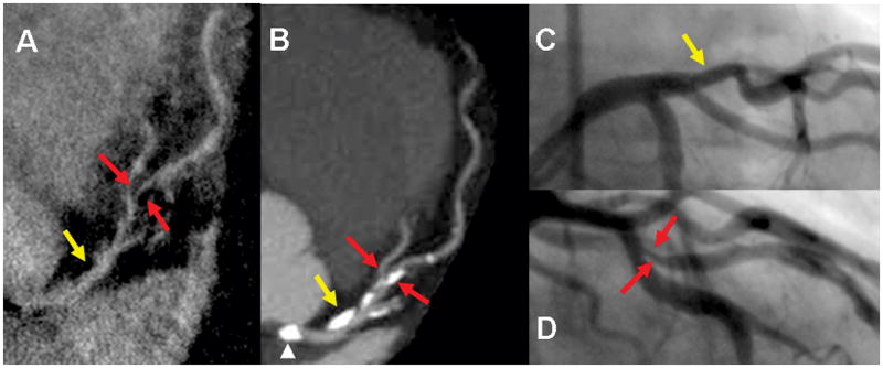
Figure 2.

Demonstrates none-significant stenosis in the proximal LAD (yellow arrow) and significant stenosis in mid LAD (red arrows) in a 76 year old male seen both on HRC (A) 320-detectors CT (B). and conventional angiogram (C and D). Despite compromised flow in high grade stenosis, signal is adequate for visualization on the HRC image. A. Multiplanar reformatted image of HRC MRA. B. maximum intensity projection (MIP) in left anterior view of the LM/LAD. C and D. Conventional angiogram of LAD in left anterior oblique views. Note calcification (arrow head) in the left cusp partially projecting over the origin of the LM on the MIP due to image plane (B) is not causing severe narrowing as seen on the conventional angiogram (C) or HRC (A)
