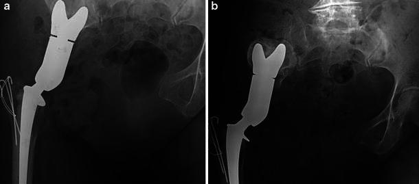
Fig. 1a–b.

X-ray films of patient P.L., 68 y.o. at the time of operation.a Postoperative AP view of the pelvis; the saddle is seated through a notch in the iliac wing.b Same view 38 months afterwards. The patient reported suffering from pain during weight bearing for the previous 2 years. Note the proximal migration of the saddle in relation to the wearing of the iliac wing
