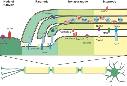
Figure 1.
Distribution of central nervous system (CNS) myelin proteins. A neuron with a myelinated axon is depicted. Myelin enwraps the axon at intervals called internodes omitting small openings termed nodes of Ranvier. Adjacent to the nodes of Ranvier are the paranode and the juxtaparanode. All four zones have a characteristic protein composition, as depicted in the upper part of the picture. At th nodes of Ranvier, Neurofascin 186 (NF186) supports the clustering of Na+ channels. To allow saltatory conduction, the nodal Na+ channels are separated from the juxtaparanodal K+ channels via the paranode, where the myelin protein Neurofascin 155 (NF155) binds tightly to the axonal complex of Contactin-1 and Contactin-associated protein (Caspr). Connexins form gap junctions between myelin layers at the paranode. 2’3’-cyclic-nucleotide 3’-phosphodiesterase (CNP) is an abundant cytoplasmic myelin protein, predominantly found at the paranode. At the juxtaparanode, clustered K+ channels are associated to Caspr-2 and Contactin-2 is found both at the innermost myelin-sheath and the axon, allowing it to bind to itself. Typical myelin proteins are found at the internode: Proteolipid protein (PLP) and Myelin basic protein (MBP) are the major myelin proteins, the quantitatively minor Myelin oligodendrocyte glycoprotein (MOG) is found at the outermost surface of the myelin sheath. Nectin-like (NECL) adhesion proteins and Myelin associated glycoprotein (MAG) are found at the periaxonal space. Nogo is also predominantly found at the adaxonal myelin membrane, its receptor Nogo-receptor 1 (NgR1) is found at the axonal membrane.

