
Figure 11.
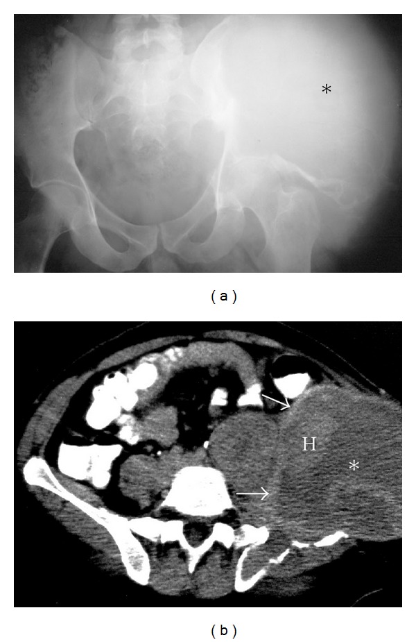
Hemophilic pseudo tumor. A 60-year-old male with hemophilic pseudo tumor of the left iliac wing. Plain radiograph (a) of the pelvis illustrating a large mass lesion (asterisk) destroying the left ilium and the acetabulum. Axial CT (b) of the pelvis demonstrates a large soft tissue density mass, centered within the left ilium (asterisk). Several areas of heterogeneous density (H) representing recent bleeds are present within the mass. Close inspection reveals a thin ballooned cortex (arrows) of the affected ilium.