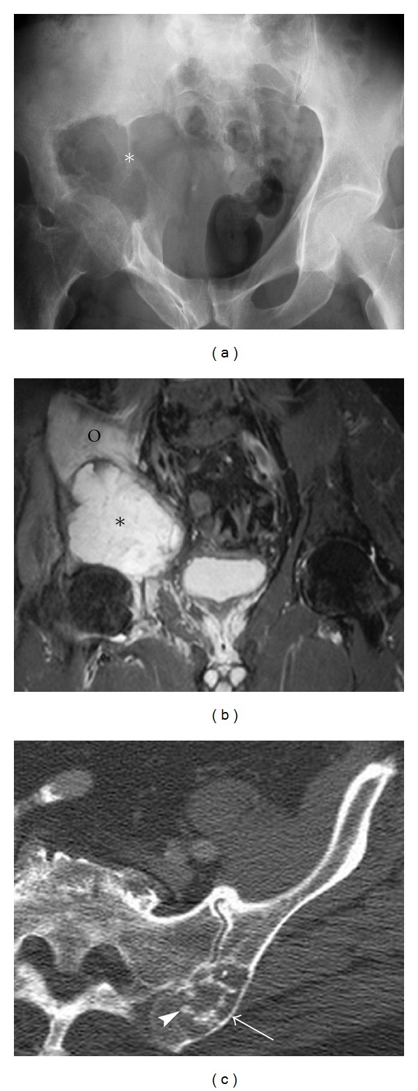
Figure 8.

Chondrosarcoma. A 62-year-old male presents with right hip pain, diagnosed with chondrosarcoma. Pelvic radiograph (a) outlines a lytic destructive abnormality centered on the superior acetabular region (asterisk). An associated soft tissue mass is seen extending into the pelvis. The relative lack of calcification of the chondroid matrix would be in keeping with a high-grade chondrosarcoma. Coronal STIR (b) demonstrates the destructive mass (asterisk) as well as associated marrow edema within the right iliac bone (circle) and a small right hip joint effusion. Axial CT scan of another patient demonstrates more typical appearances of chondrosarcoma with endosteal scalloping (arrow) and chondroid matrix (arrowhead) (c).
