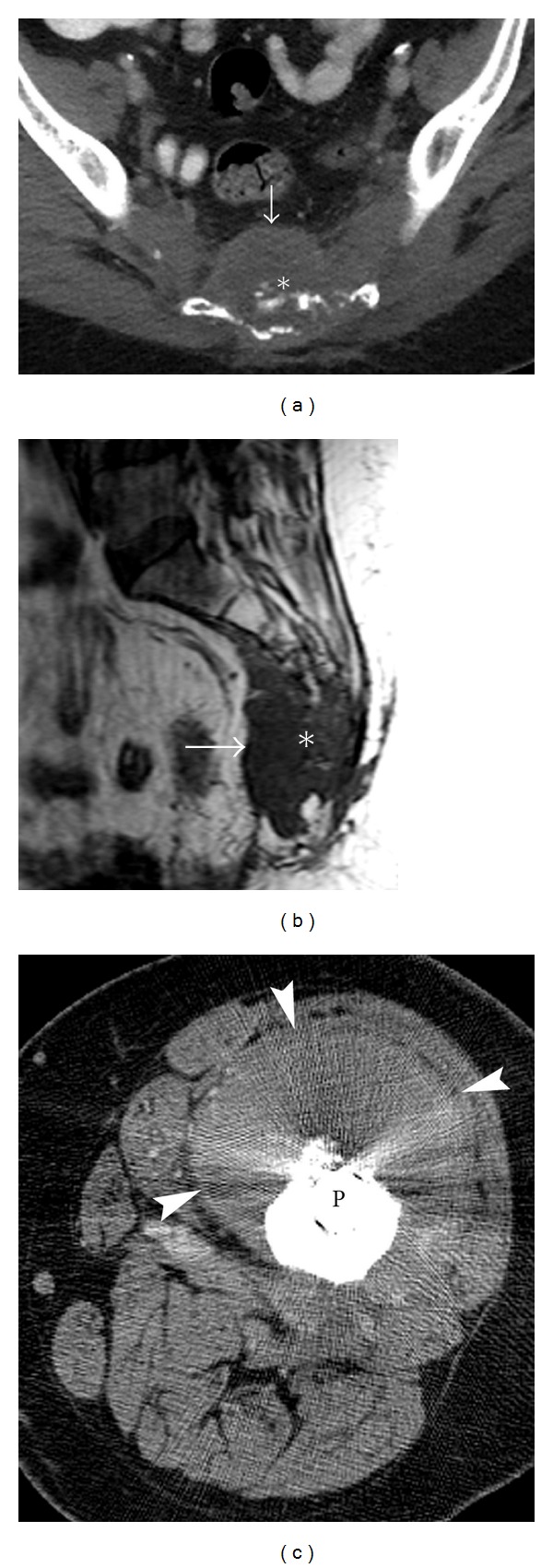
Figure 9.

Chordoma. A 64-year-old female presenting with low back pain, diagnosed with an advanced sacral chordoma. Axial CT (a) and sagittal T1 midline image (b) demonstrate a large midline mass with internal calcification, destroying the sacrum (asterisk) and infiltrating the presacral space anteriorly and epidural space posteriorly. Axial CT (c) showing a large soft tissue metastasis (arrowheads) in the thigh around the femur. Hardware artifact is from an intramedullary rod (P) internally fixing the pathological fracture of the femur.
