
Figure 3. DNPs induce systemic interferon release.
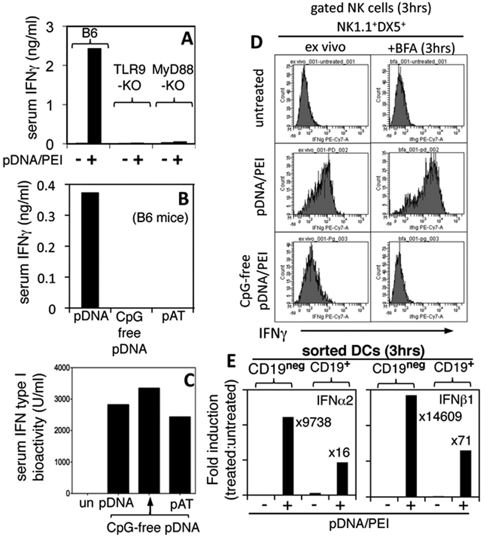
A, B. Mice were treated with DNA/PEI nanoparticles containing pDNA, CpG-free pDNA, or pAT and serum IFNγ levels were assessed after 24 hrs. (ELISA). C. Serum IFN type I levels were assessed by adding serum from DNP-treated mice to IFN bioassays (see Methods). D. Mice were treated with DNPs and after 3 hrs. splenocytes were stained with NK markers (NK1.1, DX5) and intracellular IFNγ directly (ex vivo) or after culture for 3 hr. with GolgiPlug (BFA). E. RNA from FACS-sorted CD19neg and CD19+ DCs from DNP-treated mice (3 hrs) were analyzed (qPCR) to detect IFNα2 and IFNβ1 transcripts. Data were normalized to β-actin levels in each sample and expressed as fold induction over basal levels (treated vs. untreated). Data are representative of 2 or more experiments.