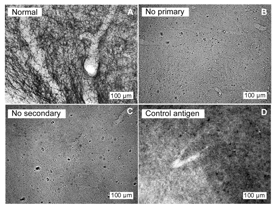
Fig. 1.
Control treatments remove SERT labeling in the IC. Sections at 20× processed in parallel from a single brain illustrating (A) normal staining of SERT fibers using primary antibody at 1:5000, (B) lack of fiber staining with omission of the primary antibody, (C) lack of fiber staining with omission of the secondary antibody, and (D) lack of fiber staining following preadsorption of the primary antibody at 1: 5000 with the control peptide. All images were normalized to the range of pixel values present.

