Figure 1.

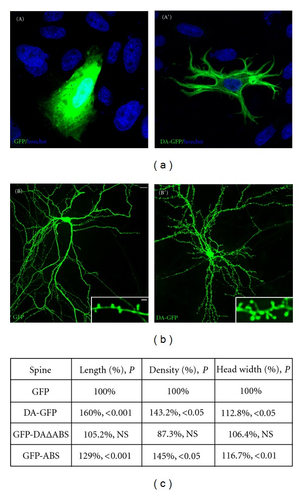
Drebrin A overexpression affects the morphology of cultured CHO-K1 cells and dendritic spines plasticity of cultured mature hippocampal neurons. In contrast to GFP (A), CHO-K1 cells transfected with DA-GFP (A′) display striking morphological changes characterized by the formation of several processes frequently branched. Blue color reveals nuclear staining by Hoechst 33258. Mature hippocampal neurons were transfected at 21 days in vitro with GFP (B) and DA-GFP (B′). After 2 days of transfection (23 days in vitro), neurons were fixed and then examined by a confocal microscope. Striking morphological changes are observed between dendritic spines of GFP- and those of DA-GFP neurons. Indeed, the dendrites of DA-GFP neurons display longer spines (inset in B′) compared with those found in GFP neurons (inset in (B)). Some spines labeled with DA-GFP can reach over 5 μm (inset in (B′), see asterisk). Scale bars: 5 μm in (A), (A′), (B), and (B′) and 2 μm in insets. (c) Table showing the spine length, density, and head width of GFP, DA-GFP, GFP-DAΔABS, and GFP-ABS neurons. GFP: green fluorescent protein; DA-GFP: drebrin A fused to GFP; GFP-DAΔABS: drebrin A without its actin binding site (ABS) fused to GFP; GFP-ABS: actin binding site of drebrin A fused to GFP; P: probability; NS: not significant.
