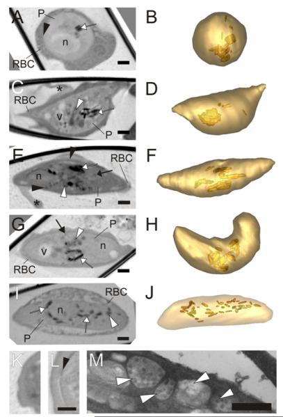
Fig.4.
Transmission X-ray tomography of sexual stages of P. falciparum. (A–J; Left panels) Individual virtual sections (64 nm) of gametocytes at stages II–V. (Right panels) Rendered models generated by segmentation of the tomograms. The parasite surface is depicted in light gold and the hemozoin crystals in deeper gold. In the tomograms the mitochondrion is indicated with a white arrow, the sub-pellicular complex with a black arrowhead, hemozoin crystals with a white arrow and dense granules (likely to be osmophillic bodies) with a black arrow. The nucleus (n), vacuoles (v) and Laveran’s bib (*) are marked. The stage II gametocyte (A and B) is roughly spherical and exhibits evidence of a sub-pellicular membrane complex (see Panel L). By stage III (C and D) formation of the crescent shape is initiated with elongation along one side. The stage IV gametocyte (E and F) is elongated with protruding extremities and shows a distinctive sub-pellicular membrane complex (black arrowhead). Some osmophillic bodies start to appear and the Laveran’s bid is evident. Stage V gametocytes (G–J) have more rounded ends and a less obvious sub-pellicular membrane complex, and prominent osmophillic bodies and mitochondrion. See Supp. Movie 1 for a translation through the tomogram shown in G. (I and J) Distribution of the hemozoin crystals in a fixed male gametocyte. (I) Virtual slice through the reconstructed tomogram of a fixed male gametocyte. (J) Segmentation model with the surface of the parasite in pale gold and the hemozoin crystals in deeper gold. (K and L) Virtual sections showing the transition between the parasite cytoplasm and the RBC cytoplasm for an asexual stage trophozoite (K) and a stage II gametocyte (L). The X-ray density at the parasite surface (arrowhead) probably represents the microtubule supported sub-pellicular membrane complex. (M) Section through a gametocyte prepared for electron microscopy. The arrowheads point to sections through the extended multi-globular mitochondrion that is present in the gametocyte. Scale bars (A–L): 1 μm; (M): 500 nm.

