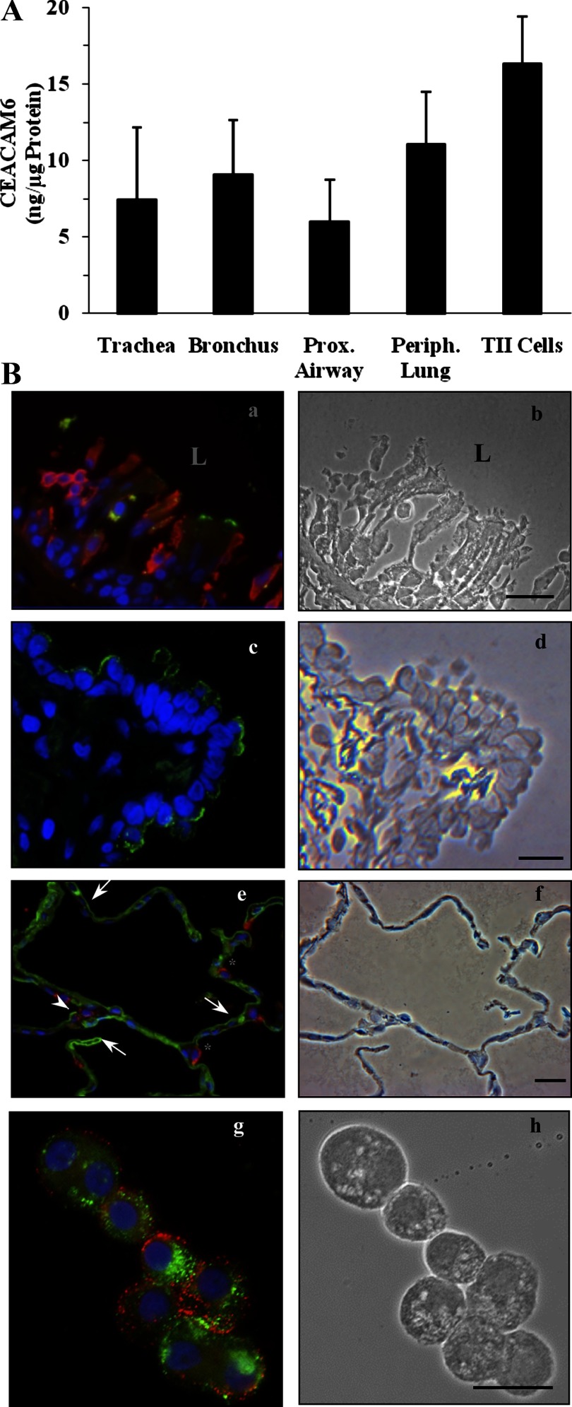
Fig. 5.
CEACAM6 expression in airway epithelium and peripheral tissue of adult human lung. A: CEACAM6 content by immunodot assay in 5 regions, proximal to distal, using scraped epithelial cells from airways, lung tissue from the periphery, and isolated type II (TII) cells. CEACAM6 is detected in all airways and alveolar cells, with no significant differences in concentrations by ANOVA. Values are means ± SE for 3–4 lungs. B: representative immunostaining. a: CEACAM6 signal (green) in some tracheal epithelial cells of a postmortem adult lung, but not in underlying interstitium. Red, cytokeratin; blue, DAPI. b: Phase image of section in a; note disrupted epithelium. L, lumen of airway. c: CEACAM6 signal (green) is detected in many, but not all, distal airway cells; this section was negative for HTII-280 [a type II cell membrane marker (red); blue, DAPI]. d: Phase image of section in c. e: Alveolar epithelium of inflation-fixed adult lung showing CEACAM6 signal (green) in epithelial cells consistent with type I cells (arrows) and in some (arrowhead), but not all, type II cells, as identified by positive SP-B (red) staining. *SP-B-positive cells that are negative for CEACAM6. f: Phase image of section in e. g: Freshly isolated type II cells with CEACAM6 signal (green) and HTII-280 (red) showing colocalization in 6 of the 7 cells in the field. h: Phase-contrast image of cells. Blue, DAPI staining. Bars = 20 μm for all images.

