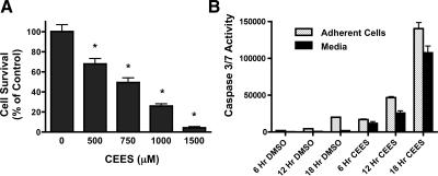
Fig. 5.
A: cytotoxicity of in vitro exposure to CEES. 16HBE cultures were exposed to diluent (DMSO alone) or diluent containing increasing concentrations of CEES (500, 750, 1,000, or 1,500 μM). After 24 h, cell viability was determined by MTT assay. Values represent means ± SD of 3 independent experiments. *Statistical difference from diluent exposure (0 μM CEES). B: effect of CEES exposure on caspase 3/7 activation. 16HBE cells were exposed to either diluent (DMSO alone) or diluent containing CEES (750 μM) for 6, 12, or 18 h. At each time point, cultures were separated into samples consisting of the adherent cell fraction (stippled bar) or the media fraction (solid bars) and tested for caspase 3/7 activity. Each bar represents average values from 9 replicate cultures exposed to CEES in 3 independent experiments. *P < 0.05 compared with diluent (DMSO) control.

