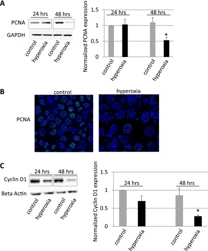
Fig. 4.
Hyperoxia downregulated proliferating cell nuclear antigen (PCNA) and Cyclin D1 expression. A: decrease in PCNA expression by Western blotting after exposure of MLE-12 cells to hyperoxia for 48 h, but not after 24 h. Densitometry analysis of 6 Western blot experiments showed a decrease of 47% in PCNA expression after 48 h hyperoxia exposure (*P < 0.05). GAPDH was used as a loading control. B: decrease in PCNA immunoreactivity (green) using confocal microscopy in MLE-12 cells exposed to hyperoxia for 48 h (magnification ×100). Nuclei were counterstained with DRAQ5 (blue). C: time-dependent decrease in Cyclin D1 expression by Western blotting and by densitometry analysis of 3 Western blot experiments (*P < 0.05). β-Actin was used a loading control. Black boxes indicate splicing of gels.

