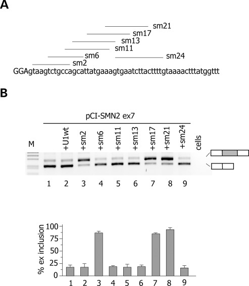
Figure 4.
Identification of ExSpeU1s in SMN2 exon 7. (A) Schematic representation of binding sites of the ExSpeU1s in SMN2 exon 7. Donor site and downstream intronic region are shown and exonic and intronic sequences are in upper and lower case, respectively. Lines correspond to the target-binding sequences of ExSpeU1s on the nascent pre-mRNA. (B) Analysis of SMN2 exon 7 spliced transcripts. The pCI-SMN2 minigene was transfected in HeLa cells alone (lane 1), with the U1wt (lane 2), or with plasmids encoding for ExSpeU1s. The pattern of splicing was evaluated by RT–PCR and amplified products were resolved on a 2% agarose gel. Lower panel is the quantification of SMN2 exon 7 splicing pattern. Data are the means ± SD of three experiments done in duplicate.

