Figure 7.
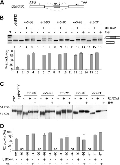
Rescue of splicing and FIX protein function by ExSpeU1. (A) Schematic representation of the pBsK-FIX minigene. Exonic sequences are boxed, introns are lines, the arrows represent primers for PCR amplification and the dotted lines indicate the two possible exon 5 alternative splicing events. The position of the ATG and TAA codons is indicated. The minigene is not drawn in scale. (B) Analysis of pBsK-FIX exon 5 spliced transcripts. pBsK-FIX exon 5 normal and mutant minigenes were transfected in BHK cells alone or with the indicated ExSpeU1s. The splicing pattern was evaluated by RT–PCR and amplified products were resolved on a 2% agarose gel. M is the molecular 1 Kb marker. Lower panel shows the quantification of the percentage exon 5 inclusion. Data are expressed as means ± SD of three independent experiments done in duplicate. (C) Western blotting of FIX in the BHK conditioned medium. PNP is pooled normal plasma. (D) FIX coagulant activity in the BHK conditioned medium. Data are expressed as means ± SD of three experiments done in duplicate.

