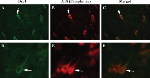
Figure 6.
Double-labeling immunofluorescence analysis of phosphorylated tau and Drp1 in AD patients. The localization of (A) Drp1 and (B) phosphorylated tau, and (C, merged) the colocalization of Drp1 and phosphorylated tau at 40× the original magnification. (D) Images of Drp1, (E) phosphorylated tau and (F) merged at 100× the original magnification. Arrows indicate localization of phosphorylated tau, Drp1 and colocalization of both.

