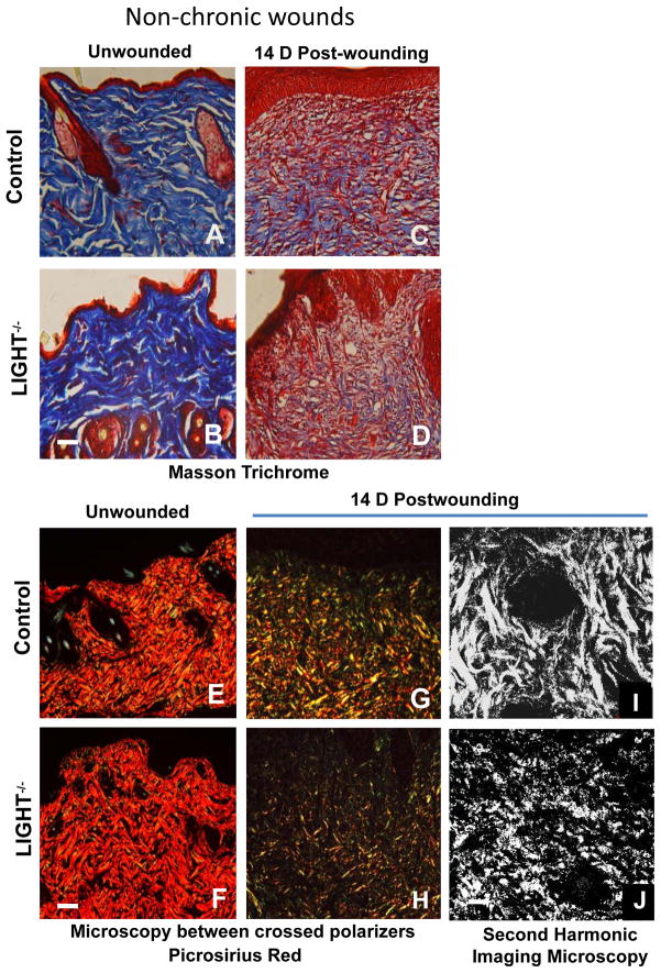
Figure 5. Collagen deposition is reduced and immature in LIGHT−/− non-chronic and chronic wounds.
(A–D) Control and LIGHT−/− unwounded skin and 14D wound tissue were stained using the Masson trichrome staining which stains the nuclei dark with Weigert’s hematoxylin, the cytoplasm and muscle fibers red with the Biebrich scarlet dye, and the collagen blue with aniline blue dye. Data shown are representative of two experiments. (E, F) Control and LIGHT−/− unwounded skin and (G,H) 14D post-wounding tissues were stained using picrosirius red, in which thick Coll I fibers appear red/orange in color and thinner Coll III fibers appear yellow/green in standard 10μm thick tissue sections viewed between crossed polarizers. Scale bar = 50μm. (I,J) Second-harmonic imaging microscopy (SHIM) of the structure of Coll I fibers in 14D wild-type and LIGHT−/− wounds Scale bar = 100μm. Data shown are representative of two independent experiments.

