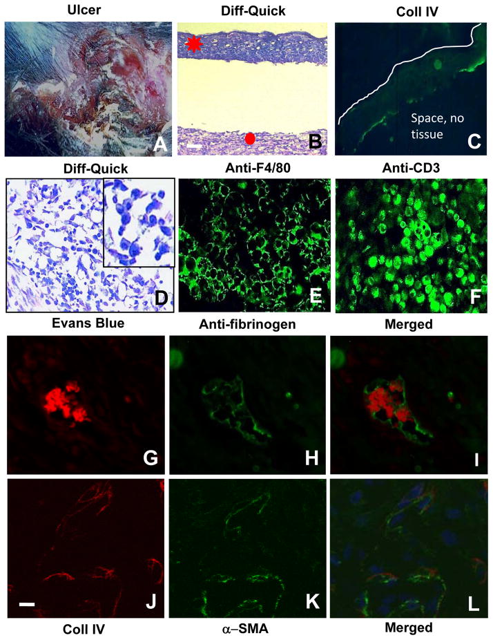
Figure 7. Defects in the LIGHT−/− mice chronic wounds are exacerbated when compared with non-chronic wounds.
(A) General appearance of the chronic wounds from the LIGHT−/− mice. (B) Highly impaired epithelial-dermal connectivity. (C) Chronic wound epidermis is immature; immunolabeling for Coll IV shows a discontinuous epithelial basement membrane (green). Below the staining there is an empty space much like in B. Line delineates the top of the epidermis (D) Histological examination showed the presence of large numbers of small round and dense cells, likely inflammatory cells; insert show enlargement of the cells). (E–F) Immunolabeling of macrophages with anti-F4/80 (E) and T-lymphocytes with anti-CD3 (F) in the chronic wounds. (G–I) Chronic LIGHT−/− wound tissue was labeled with Evans blue (red fluorescence) and anti-fibrinogen antibody (green fluorescence) to detect intravascular coagulation. (J–L) Co-staining with antibodies against Coll IV (red) and αSMA (green) in chronic LIGHT−/− wounds shows very low levels of both. Blue fluorescence is DAPI. Scale bar for B–D = 50μm for E–L = 20μm. Data shown are representative of staining of multiple sections of the chronic wounds.

