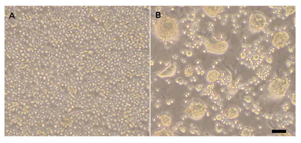
Figure 1.
Phase contrast photomicrographs of control and infected C6/36 cells after infection. (A) Control cells. (B) Cells 3 days after infection with SDDM06-11. Note the extensive cell fusion and syncytia formation in the infected cells. Microscope settings Ocular: 10; Lens: 10X. Scale bar, 50 μm.

