Figure 1.
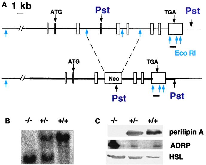
Generation of the peri null mouse. (A) Targeted mutation of the murine peri gene. Upper diagram indicates the nine exons in boxes of the peri gene, with translation start and stop sites for the predominant Peri A form (3) and EcoRI and PstI sites. Below is the mutated peri gene with the Neomycin resistance cassette inserted into the EcoRI sites of introns 3 and 6. The position of the insertion would disrupt coding of all four peri mRNA species(X. Lu, J.G.-G., N. G. Copeland, D. J. Gilbert, N. A. Jenkins, C.L. & A.R.K., unpublished work). The region used for homologous recombination is indicated by the thick line. The bar below exon 9 represents the position of the downstream probe used to assess homologous recombination within the peri locus in PstI digests for genomic Southern blots. (B) Southern blotting of tail DNA digested with PstI. (C) Immunoblotting of adipose tissue extracts for peri A, ADRP, and HSL. Adipose tissue samples were extracted and proteins solubilized as described under Experimental Procedures. Gel lanes were loaded with the equivalent of proteins extracted from 10 mg of adipose tissue.

