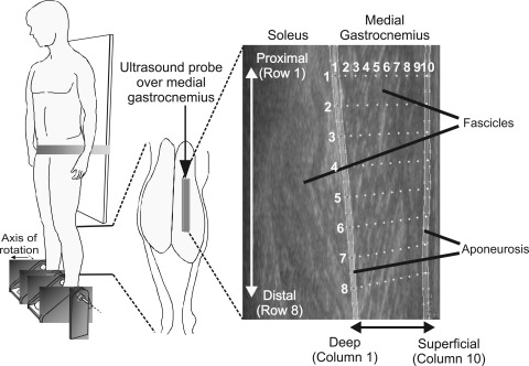
Fig. 1.
Schematic representation of setup used to collect ultrasound images. Participant stood on two footplates, supported by a waistband wrapped around a fixed vertical board. Ultrasound probe (grey block) was placed over the midbelly region of the medial gastrocnemius (MG) muscle. Ultrasound images reveal the MG and soleus muscles, with superficial corresponding to the region closest to the skin and deep corresponding to the region of muscle closest to the tibia. Configuration of probes, automatically placed on the first image of a sequence, within the segmented regions of medial gastrocnemius is also shown (vertical lines and small points, see methods for full explanation). Initial configuration was a 10 × 8 grid (column and row numbers denoted in white). For comparison of tracked muscle movements using the cross-correlation approach (13), templates were manually placed along the deep and superficial aponeurosis in the most representative frame. Templates across the fascicle region were then positioned using interpolation, providing the same configuration shown.

