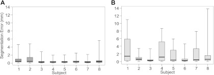
Fig. B1.
Box and whisker plots showing the error of the segmentation using ASM* (A) and ASM (B) in each participant. Error was the absolute distance between the manually and ASM*/ASM defined position of each landmark. Plots show the error calculated from each landmark within each analyzed image (total 76 landmarks × 75 frames = 5,700 points in each box). Middle bar represents the median value, bottom and top of the box represent the 25th and 75th percentiles, respectively, and the whiskers represent the minimum and maximum values.

