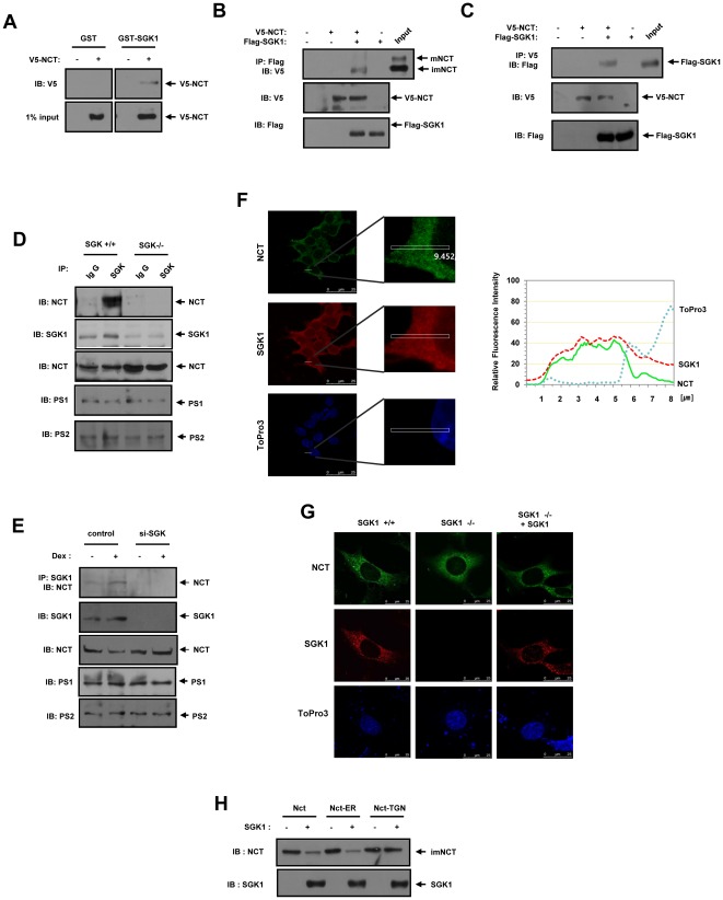
Figure 5. NCT interacts directly with SGK1 in intact cells.
(A) Recombinant GST or GST–SGK1 proteins were immobilized onto GSH-agarose. HEK293 cells were transfected with an expression vector encoding for 2 µg of V5-NCT or an empty vector. After 48 hours of transfection, the cell lysates were subjected to GST pull-down experiments with immobilized GST or GST–SGK1. Proteins bound to GST or GST–SGK1 was analyzed via immunoblotting with an anti-V5 antibody. The input represents 1% of the cell lysate prior to in vitro binding assay. (B, C) HEK293 cells were transfected with expression vectors encoding for 2 µg of V5-NCT and 2 µg of Flag-SGK1 as indicated. (B) After 48 hours, the cell lysates were subjected to immunoprecipitation (IP) with anti-Flag. The immunoprecipitates were then immunoblotted (IB) with anti-V5. (C) Cell lysates were subjected to immunoprecipitation with an anti-V5 antibody, and the immunoprecipitates were immunoblotted with an anti-Flag antibody. (D) MEF cells from SGK1+/+ and SGK1−/− mice were lysed and subjected to immunoprecipitation with immunoglobulin G (IgG) and anti-SGK1 antibodies as indicated. Immunoprecipitates were immunoblotted with an anti-NCT antibody. (E) Rat fibroblast Rat2 cells expressing either control siRNA or rSGK1 siRNA were left untreated or treated with 1 µM dexamethasone for 24 hours. Rat fibroblast Rat2 cells expressing either control siRNA or rSGK1 siRNA were lysed and subjected to immunoprecipitation with anti-SGK antibody. Immunoprecipitates were immunoblotted with an anti-NCT antibody. (F, G) MEF from SGK1+/+, SGK1−/− mice or HEK293 cells or were stained with Alexa 488 (green) and Alexa 546 (red) and examined by confocal microscopy. The DNA dye ToPro3 was used to visualize nuclei of all cells. For each experiment, at least 200 cells were examined, and the figures shown here represent the typical staining pattern for a majority of cells and quantify the fold enrichment at the indicated region (white box). (H) HEK293 cells were transfected for 48 hours with expression vectors encoding for 1 µg of NCT, NCT-ER, NCT-TGN and 2 µg of Flag-SGK1. The cell lysates were immunoblotted with anti-Flag, and anti-NCT antibodies. (A–E, H) The cell lysates were also subjected to immunoblotting analysis with the indicated antibodies. These results represent one of three independent experiments. IB, Immunoblot. IP, Immunoprecipitation.

