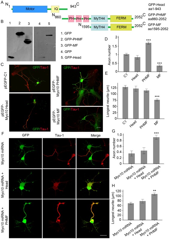
Figure 4. Overexpression of Myo10 PHMF induces the formation of multiple axon-like neurites.
A, Schematic representations of Myo10 Head, Myo10 PHMF and Myo10 MF. B, Western blot detects expression of GFP, GFP-Head, GFP-PHMF and GFP-MF. C, Neurons transfected with pEGFP-C1, pEGFP-Myo10 Head, pEGFP-Myo10 PHMF and pEGFP-Myo10 MF respectively were labeled with anti-Tau-1 antibody at DIV 3. D and E, Quantitative analysis of average number of axons and the longest neurites. F, Myo10 PHMF is sufficient for axon formation. Neurons transfected with Myo10 miRNA together with Myo10 Head and Myo10 PHMF respectively were stained with Tau-1 antibody at DIV 3. G and H, Quantitative analysis of average number of axons and average length of the longest neurites. Scale bar, 20 µm. **P<0.01; ***P<0.001.

