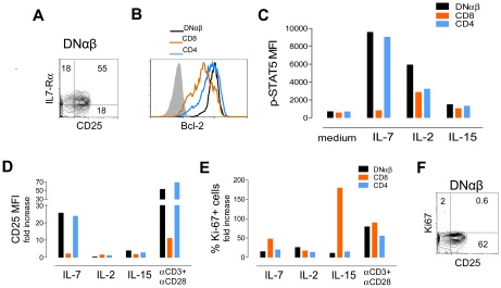
Figure 5. Ability of expanded circulating DNαβ T-cells to respond to IL-7 and IL-2.
Representative flow cytometric analysis of freshly isolated PBMC from a patient with R255X FOXN1 mutation 59 months after thymic transplantation illustrating (A) the preserved expression of IL-7Rα and increased CD25 expression within DNαβ T-cells; and (B) the high levels of Bcl-2 expression within DNαβ in comparison with CD8 and CD4 T-cells. (C) Up-regulation of p-STAT5 upon 15 min stimulation of PBMC with IL-7 (50 ng/ml), IL-2 (100 U/mL) or IL-15 (25 ng/ml), bars represent p-STAT5 MFI within gated DNαβ, CD8 and CD4 T-cells. Freshly isolated PBMC were cultured for 5-day in the presence of IL-7 (10 ng/ml), IL-2 (10 U/mL), or IL-15 (12.5 ng/ml) or anti-CD3 plus anti-CD28 stimulation and graphs represent the fold change of CD25MFI with respect to medium (D), and the frequency of Ki-67+ cells (E), within gated DNαβ, CD8 and CD4 T-cells. (F) Representative analysis of freshly isolated PBMC illustrating the low levels cycling cells (Ki-67+) despite the increased CD25 expression within gated DNαβ T-cells. Numbers inside dot-plots represent frequency of cells expressing the indicated molecules acquired with FACSCanto (p-STAT5 and 5-day cultures) or FACSCalibur flow cytometers.

